Our services
We are passionate about engaging you in your cardiovascular health. We believe that good heart health begins at home, and we strive to make your experience in our clinic a memorable, pleasant experience combining the best in cutting-edge treatment options and therapies along with a comprehensive education about how you can take control of your own life, reduce the risk of heart disease, diabetes and stroke, and change the way you live. We will help you!
Cardiovascular clinic
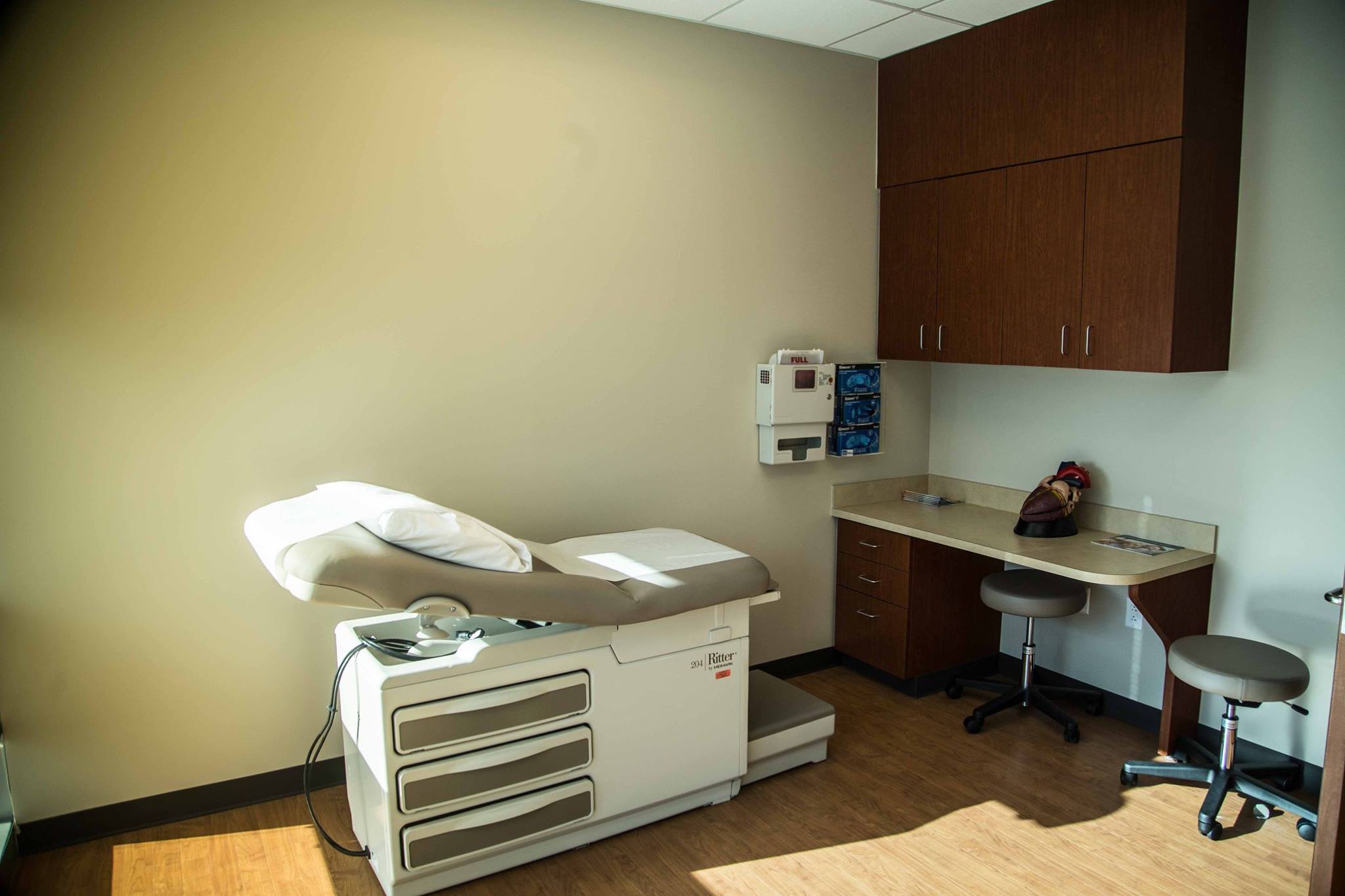
We are Houston Methodist Cardiology Associates. In affiliation with Houston Methodist Sugar Land Hospital, the largest community hospital of the Houston Methodist System and the largest hospital in Fort Bend County, we have a state-of-the-art cardiology clinic, with multiple facilities at your service. Our clinic is located on 3527 Town Center Boulevard South, in Sugar Land, Texas, across the street from Houston Methodist Sugar Land Hospital’s Emergency entrance.

Electrocardiogram (EKG/ECG)
An Electrocardiogram (abbreviated EKG or ECG) is a test where your cardiologist will measure the electrical signals in the heart. Like any other machine, the heart has its own electrical conduction system that conducts signals between its different components. The EKG is designed to be very sensitive to the flow of these signals through the heart to help diagnose two types of problems. First, when there is any damage to your heart muscle because of a heart attack (see heart attack) the flow of electricity is disturbed by the damaged heart tissue, and this can be detected. Second, the electrical system of the heart is also the component that controls the precise timing of the heart rhythm. When there is a disturbance in the heart rhythm (called arrhythmia), this abnormal electrical conduction can also be seen on the EKG. All of our EKGs are performed and interpreted right within our office(what should I expect?)
Stress Tests
A stress test is designed to assess two things, often administered in two parts. The first part is the stress component. Here, we will evaluate whether you have good functional capacity, which is a measure of how well your heart can perform. This assessment is made by asking you to walk on a treadmill. Cardiologists employ a specific program called the "Bruce Protocol" to assess how well you can tolerate vigorous exercise in a step-wise fashion. Sometimes, if you cannot run on a treadmill (leg pain, knee problems, foot injury, etc), we have an alternative way to stress your heart by using a chemical stress test. In this test, we will administer a chemical that will cause your blood vessels to increase in size temporarily and allow more blood to flow into the heart. This is similar to what your heart would experience if you were on a treadmill. The second part of the test is the perfusion component. In this portion of the test, you will be given an injection containing a radiotracer chemical which incorporates into the heart muscle temporarily. The radiotracer's distribution in your heart is proportional to the blood flow into various parts of your heart. This distribution can be monitored by taking pictures of your heart using a specialized camera called a "nuclear camera". Our stress tests are performed directly in the office setting. (What should I expect?)
TRANSTHORACIC echocardiograms (ECHO)
A Transthoracic echocardiogram is like taking a photograph of your heart, except that it is more like filming your heart in action. During this procedure, you will lie on a flat surface, and our echo technologist will place a probe over your chest to capture still frames and video clips of your heart in action. The probe tip has a special camera that can see through your skin, bones and muscle and show us your heart as it is working inside your body. It's completely non-invasive and involves no pain, no anesthesia, no sedation. Remember how a baby's sonogram is assessed when someone is pregnant? Well, this is the same technology, except that instead of a baby, your heart is being imaged. Echocardiograms (often abbreviated as "echo"), give cardiologists a lot of information about your heart. It tells us whether your heart is squeezing well, whether all the valves are working well, how the blood flow goes through your heart and whether there is any fluid around your heart. All of our echoes are performed right in our office. (what should I expect?)
transesophageal echocardiograms (T.E.E.)
A transesophageal echocardiogram (referred to commonly as "T.E.E.") is a more invasive version of the regular echo. This procedure is done within the hospital, often in an outpatient setting (meaning, you come into the hospital in the morning, have the procedure done, and leave in the afternoon). In this procedure, a small, flexible, and slender probe is "swallowed" by the patient. The probe transits in the esophagus (food pipe) and transmits images of your heart. The back of your heart sits directly in front of your esophagus, so the close proximity of the echo probe to the heart means that your cardiologist can capture spectacular looking, high resolution images of your heart from this location. When we are searching specifically for things that require precise accuracy (for example, blood clots in the heart, looking at the structure of valve leaflets, looking for small bacterial growths in the heart valves etc), the high resolution fidelity of a T.E.E. is a must. Since the probe has to be swallowed, this can be uncomfortable, and to mitigate that discomfort we provide moderate sedation (not general anesthesia) to the patient. Our T.E.E. studies are done in the Memorial Hermann Sugar Land Hospital. (what should I expect?).
cardiac rhythm monitoring (holter)
Cardiac Rhythm Monitoring services are provided for patients with suspected rhythm abnormalities. If you are suspected of having an arrhythmia (abnormal heart rhythm) (see arrhythmia), this service is generally for you. We will fit you with a wearable device that will monitor your heart rate, rhythm and transmit it to a remote facility. This service can be done in one of three ways. The first is a hands-off monitor that will record your heart rhythm without your input, called a Holter Monitor. This comes in 24 hr and 48 hr monitoring periods, depending on the frequency of your rhythm disturbance. The second is a patient-input monitor called an Event Monitor. This is designed to monitor your heart rhythm closely when you trigger the device by pressing a button. This is usually fitted for 30 days, for patients whose rhythm abnormalities are more rare. The third, rarer device is called an Implantable Loop Recorder (ILR), which is implantable and delivered under the skin of your chest wall via a small incision in the office. The ILR can remain in your body for upto 2 years completely safely, and then removed via a similar small in-office procedure. The All of these are done within the comfort of our clinic. (what should I expect?).
cardiac catheterization
When your cardiologist strongly suspects that you have coronary artery disease, or blockages in the blood vessels of your heart, you will be scheduled for a cardiac catheterization. This procedure often has many parts, including the option to place a stent to open a blocked artery if one is found at the time. This is an invasive procedure that is usually done within the cardiac catheterization lab (or a specialized room) within the Houston Methodist Sugar Land Hospital. During this procedure, your cardiologist will place small plastic tube into your artery usually in your leg or in your wrist, and thread a small flexible plastic tube (called a "catheter") through the artery all the way into your heart under X-ray guidance. Once in the heart, the cardiologist will enter the opening of the coronary arteries, the arteries that can be blocked during heart disease and inject contrast dye into these vessels. The flow of contrast dye through the artery can be captured on X-ray video and it shows whether the artery is open, narrowed or completely closed giving options for treatment. If no treatment is performed, patients can generally go home the same day. If the patient is treated with a stent, then the patient generally stays overnight. Please read more in detail about cardiac catheterization. If you'd like more information about your procedure with us, read what should I expect?.
PERIPHERAL ANGIOGRAPHY
In addition to correcting blocked arteries of the heart, we also specialize in fixing blockages in the arteries of the leg. The procedure for correcting this problem is called a peripheral angiogram. In this procedure, your cardiologist will place a small plastic tube into the artery in your leg, and inject contrast dye through it directly to look at the blood flow in the affected leg. If there is a blockage, it can be corrected quite readily with the use of balloons and catheters, or with the use of specialized stents to restore blood flow to the leg. Please read about this procedure in detail under peripheral vascular disease. Also read what should I expect?